

|
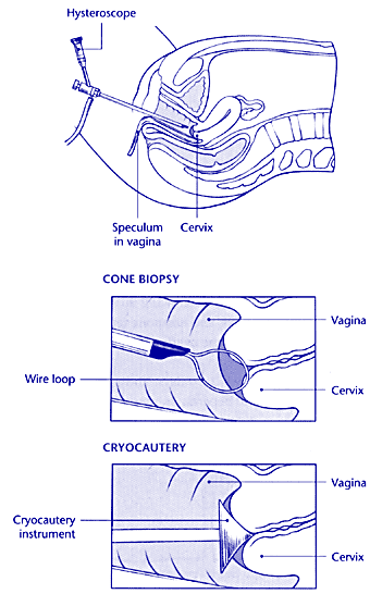
Cone biopsy and cryocautery of the cervix • Cone biopsy and cryocautery of the cervix are two methods used to remove abnormal cervical cells. • Cone biopsy may be performed in out-patients under local anaesthetic or as a day-case operation in theatre under general anaesthetic. Cryocautery is a painless procedure carried out in out-patients. • In both procedures, an instrument called a speculum
is inserted to hold the walls of the vagina apart and the
cervix ª For a cone biopsy, the area to be removed is highlighted
using iodine solution. A 'cone' of tissue, containing the
abnormal area, is then removed using either a hot, wire
• For cryocautery, the abnormal cells are destroyed
by • There may be some slight bleeding following these
procedures. A vaginal discharge, which is dark brown • Sexual intercourse should be avoided until after the next menstrual period to ensure that the cervix heals properly. • Normal activities can he resumed the following day. |
 |