|
Vulval biopsy and treatment of VIN
• To diagnose vulval intraepithelial neoplasia (VIN for short),
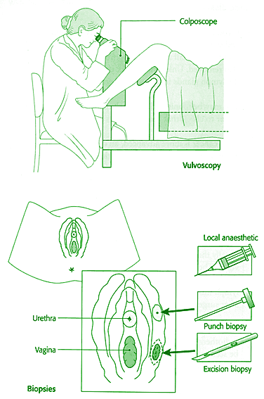
the vulva is examined in a procedure called vulvoscopy. A special microscope
called a colposcope is usually used. Vulvoscopy is a painless, out-patient
procedure. You will be asked to lie on your back with your legs in supports.
• Dilute acetic acid may be painted onto your vulva to show up
any abnormal cells. This is not painful, but may cause mild irritation.
• Samples of vulval tissue, called vulval biopsies, may be removed
for examination in the laboratory. These are taken either after numbing
the area with local anaesthetic, or under a general anaesthetic. Your
doctor may take several small circular punch biopsies (tiny circular
samples of skin), or remove a larger piece of tissue (called an excision
biopsy). You will probably need a few stitches after an excision biopsy,
and these will leave a small scar.
• If you have VIN, the abnormal cells may be removed, or sometimes
destroyed using a laser. This is usually carried out under general anaesthetic.
You will be given painkillers and possibly also a local anaesthetic
jelly to use for a few days after treatment.
• After VIN has been diagnosed, you will require regular vulvoscopy.
An alternative way of managing this condition is to remove only areas
that appear to be progressing towards early cancer. This may save unnecessary
surgery.
|
|
 |